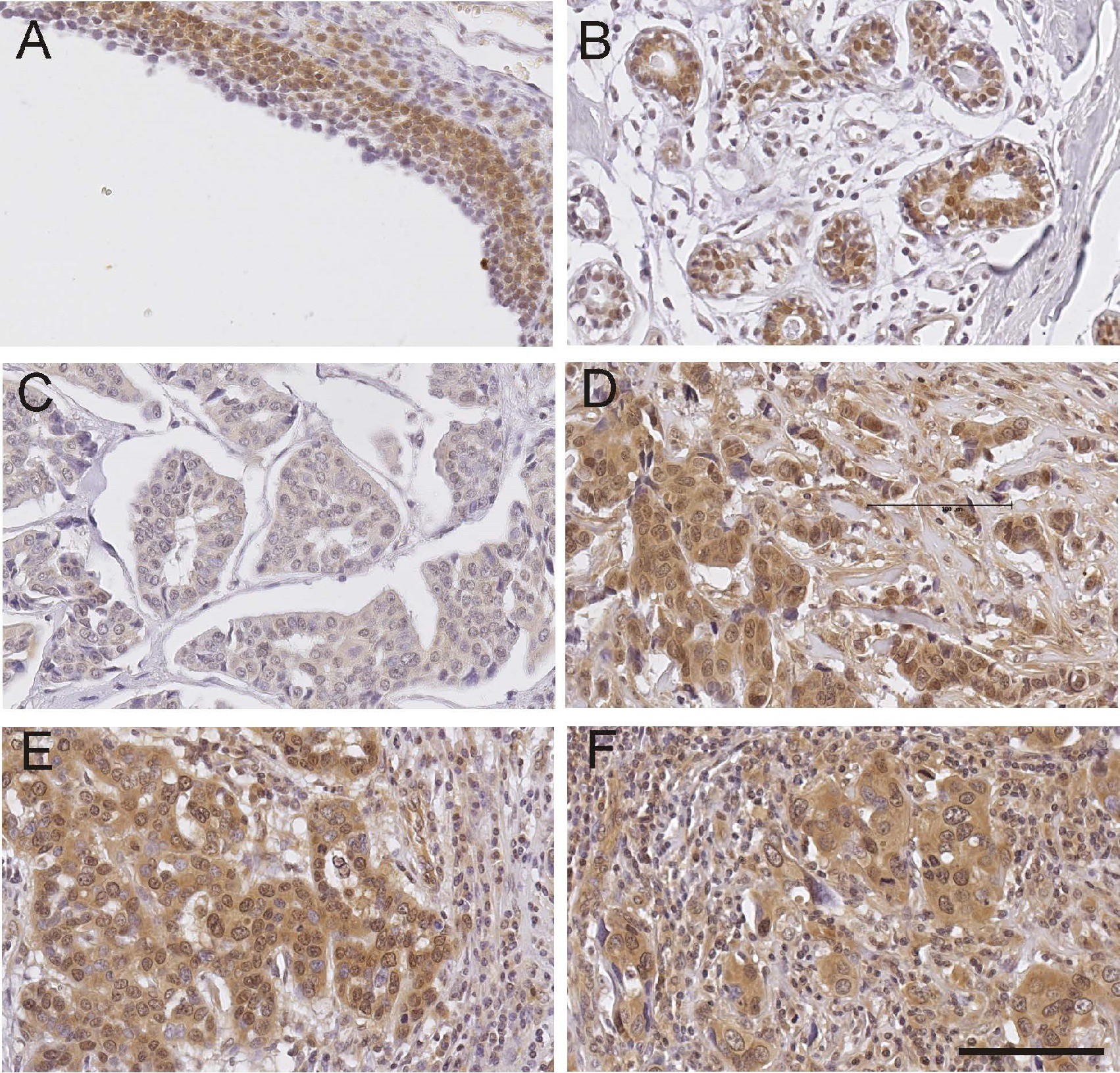
Fig. 1. ADAMTS1 immunohistochemistry. a) normal ovary, b) normal breast tissue, c) breast cancer tissue with low ADAMTS1 immunostaining, d) breast cancer tissue with high ADAMTS1 immunostaining. e) and f) are matching primary and metastatic breast cancer tissues from the same patient. scale bar = 100µm, all images are the same magnification